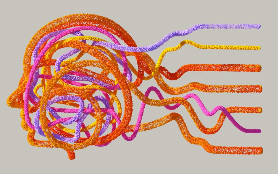
Hidden Layers in the Brain's Memory Center: A New Blueprint for Understanding Memory and Disease
A groundbreaking study from the Keck School of Medicine of USC has revealed a previously unknown four-layer structure within the hippocampal CA1 region, a critical hub for memory, navigation, and emotion. Using advanced RNA imaging to map over 330,000 genetic signals, researchers discovered distinct, shifting bands of neuron types. This new architectural blueprint transforms our understanding of how different parts of the hippocampus support specific behaviors and offers crucial insights into why certain neurons are more vulnerable in disorders like Alzheimer's disease and epilepsy.
For decades, the hippocampus has been recognized as the brain's central command center for memory formation, spatial navigation, and emotional processing. Yet, the precise internal architecture of one of its key subregions, CA1, has remained elusive. A landmark discovery, published in Nature Communications by researchers at the Mark and Mary Stevens Neuroimaging and Informatics Institute, has now lifted the veil, revealing a sophisticated four-layer blueprint hidden within this vital structure. This finding, made possible by cutting-edge molecular mapping, is poised to reshape our fundamental understanding of brain organization and its breakdown in neurological diseases.

The Discovery: Mapping the Brain's Hidden Architecture
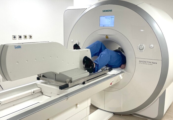
The research team, led by senior author Michael S. Bienkowski, PhD, employed a high-resolution RNA labeling technique called RNAscope. This method allowed them to visualize gene expression at the single-molecule level within tens of thousands of neurons in the mouse hippocampus. By analyzing more than 330,000 RNA signals from over 58,000 CA1 pyramidal cells, they constructed an unprecedented cellular atlas.
The data revealed a stunning pattern: instead of a blended mosaic, the CA1 region is organized into four thin, continuous layers of specialized cell types. Each layer is defined by a unique molecular signature—a specific pattern of active genes. As co-first author Maricarmen Pachicano described, visualizing these patterns showed "clear stripes, like geological layers in rock." Crucially, these layers are not static; they subtly shift in thickness and shape along the length of the hippocampus, meaning different segments of CA1 contain distinct mixtures of neuron types.
Implications for Memory, Behavior, and Disease
This layered organization provides a powerful new framework for explaining brain function. Researchers have long suspected that different parts of the CA1 support different aspects of learning and memory. This discovery offers the physical substrate for that theory. The specific blend of neuron types in a given location likely determines its functional role, whether in forming a specific memory, processing spatial information, or influencing emotional state.
Perhaps most significantly, the finding sheds new light on neurological disorders. The hippocampus is among the first regions affected in Alzheimer's disease and is heavily implicated in epilepsy. The layered model suggests that diseases may target specific cell types within a particular layer. "If a disease targets one layer's cell type, the effects will vary depending on where in CA1 that layer is most prominent," explained Bienkowski. This could explain the selective vulnerability of certain neurons and the varied progression of symptoms in these conditions.
A New Tool for Global Neuroscience
Beyond the conceptual breakthrough, the team has created a tangible resource for the scientific community. They compiled their findings into a new, freely available CA1 cell-type atlas as part of the Hippocampus Gene Expression Atlas (HGEA). This resource includes interactive 3D visualizations accessible through an augmented-reality app, allowing researchers worldwide to explore this intricate structure in detail.
While the study was conducted in mice, the researchers note that similar layered arrangements have been observed in primates. The fundamental organization may be a shared feature across mammalian species, making this discovery a critical stepping stone for understanding the human brain. As Arthur W. Toga, PhD, director of the Stevens INI, stated, "Discoveries like this exemplify how modern imaging and data science can transform our view of brain anatomy."

The Path Forward
The identification of these hidden layers opens numerous avenues for future research. The immediate next step, as Bienkowski notes, is to "understand how these layers connect to behavior." Scientists now have a precise anatomical framework to investigate how specific neuron layers contribute to memory, navigation, and emotion. Furthermore, this blueprint will guide translational studies aimed at developing targeted therapies for Alzheimer's, epilepsy, and other conditions by focusing on the most vulnerable cell populations.
This discovery underscores a profound shift in neuroscience: moving from viewing brain regions as uniform tissues to understanding them as precisely organized, layered circuits with specialized components. It reaffirms that to decipher the brain's most complex functions—and its failures—we must first map its most fundamental structures.