Zap-and-Freeze Imaging: A New Window into Brain Communication and Parkinson's Disease
A groundbreaking 'zap-and-freeze' imaging technique developed by Johns Hopkins researchers is providing unprecedented views of how brain cells communicate. By freezing neural tissue at the precise moment a signal fires, scientists have captured the rapid behavior of synaptic vesicles in both mouse and human neurons. This research offers crucial insights into the mechanisms of sporadic Parkinson's disease, which accounts for most cases without inherited genetic causes. The findings, published in Neuron, reveal conserved molecular processes across species and open new pathways for understanding neurodegenerative disorders and developing potential therapies.
Neuroscience has long faced a fundamental challenge: brain cells communicate at speeds too rapid for conventional imaging techniques to capture. This limitation has hindered our understanding of neurological disorders, particularly those like Parkinson's disease that involve subtle disruptions in synaptic transmission. A revolutionary imaging breakthrough from Johns Hopkins Medicine is now changing this landscape. Using a high-speed "zap-and-freeze" method, researchers have achieved their clearest view yet of how neurons exchange messages, offering new clues about the origins of nonheritable Parkinson's disease and potentially guiding future therapeutic development.

The Zap-and-Freeze Technique Explained
The zap-and-freeze method represents a significant advancement in cellular imaging technology. As described in the Johns Hopkins research, the technique involves delivering a brief electrical stimulus to activate brain tissue, followed immediately by rapid freezing that preserves cellular structures in their exact positions at the moment of signal transmission. This approach allows researchers to capture events that typically unfold in milliseconds—far too quickly for traditional microscopy methods. The preserved tissue can then be examined in detail using electron microscopy, revealing the precise arrangement of synaptic components during neural communication.
This methodology builds on earlier work published in Nature Neuroscience, where researchers first demonstrated its potential for visualizing fast changes in synaptic membranes. The current study, supported by the National Institutes of Health and published in Neuron on November 24, 2025, represents the technique's application to both mouse and human brain tissue. By comparing these two systems, researchers can validate whether observations in animal models accurately reflect human neurobiology—a critical consideration for translating basic research into clinical applications.
Insights into Synaptic Vesicle Dynamics
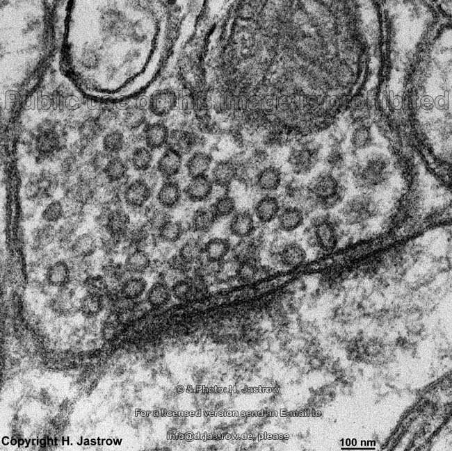
At the heart of neural communication are synaptic vesicles—tiny packages that carry chemical messengers (neurotransmitters) from one neuron to the next. In a healthy brain, these vesicles undergo a carefully orchestrated cycle of release, retrieval, and recycling. The zap-and-freeze technique has revealed this process in unprecedented detail, capturing the moment when vesicles fuse with the cell membrane to release their contents and the subsequent retrieval through endocytosis.
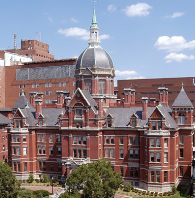
The research team, led by senior author Shigeki Watanabe, Ph.D., applied the technique to living cortical brain tissue obtained from six patients undergoing epilepsy surgery at The Johns Hopkins Hospital. These samples, collected with permission during procedures to remove hippocampal lesions, provided rare access to viable human neural tissue. Collaborating with researchers from Leipzig University in Germany, the team first validated the method in mouse tissue by observing calcium signaling—the trigger that prompts neurotransmitter release.
When applied to human tissue, the researchers documented the same vesicle recycling steps occurring in human neurons. Particularly significant was the identification of Dynamin1xA—a protein essential for ultrafast synaptic membrane recycling—at locations where endocytosis occurs in both species. This conservation of molecular mechanisms between mice and humans strengthens the validity of using mouse models to study human brain biology and disease processes.
Implications for Parkinson's Disease Research
Parkinson's disease represents one of the most common neurodegenerative disorders, with sporadic cases accounting for the majority of diagnoses according to the Parkinson's Foundation. These nonheritable forms involve disruptions at the synapse—the tiny junction where neurons communicate. Because synaptic activity happens so rapidly and at such small scales, it has been exceptionally challenging to study in the context of disease.
The zap-and-freeze technique offers new possibilities for understanding these disruptions. Watanabe notes that visualizing synaptic membrane dynamics in live brain tissue samples may help researchers understand similarities and differences between nonheritable and heritable forms of Parkinson's. The ability to observe vesicle behavior in real-time (or more accurately, at the moment of freezing) could reveal how communication begins to fail in affected neurons.
Looking forward, the research team plans to apply the zap-and-freeze method to brain tissue collected from individuals with Parkinson's disease who are undergoing deep brain stimulation procedures. This comparative approach could identify specific alterations in vesicle dynamics that characterize the disease state. Such insights might eventually guide the development of targeted therapies that address synaptic dysfunction rather than merely managing symptoms.
Broader Scientific and Therapeutic Implications
Beyond Parkinson's disease, the zap-and-freeze technique has broader implications for neuroscience and medicine. The ability to capture rapid synaptic events could advance our understanding of other neurological and psychiatric conditions where synaptic communication is disrupted, including epilepsy, Alzheimer's disease, and various neurodevelopmental disorders. The technique's validation in human tissue is particularly significant, as it bridges the gap between animal research and human biology.
The research was supported by multiple funding sources including the National Institutes of Health, Howard Hughes Medical Institute, and the Chan Zuckerberg Initiative, reflecting the broad scientific interest in this methodological advancement. As Watanabe emphasizes, the technique could eventually guide therapy development for neurodegenerative disorders by providing a more detailed understanding of where and how synaptic communication fails.
For the neuroscience community, the study demonstrates the power of combining innovative imaging techniques with access to human tissue samples. The collaboration between Johns Hopkins Medicine and Leipzig University highlights the international nature of cutting-edge brain research and the importance of shared methodologies for advancing our understanding of human neurobiology.
Conclusion
The zap-and-freeze imaging technique represents a significant leap forward in our ability to study brain communication at the cellular level. By capturing synaptic events that were previously too rapid to observe, researchers have gained new insights into the fundamental mechanisms of neural transmission and how these processes might be disrupted in Parkinson's disease. The conservation of these mechanisms between mice and humans validates animal models while opening new avenues for human-specific research.
As this technology continues to develop and is applied to disease states, it promises to deepen our understanding of neurodegenerative disorders and potentially identify new therapeutic targets. The journey from basic imaging technique to clinical application will require further research, but the zap-and-freeze method has already provided neuroscience with a powerful new tool for exploring the intricate world of synaptic communication.